
Communicated by the Basic and Translational Myology Team
Alterations of intestinal smooth muscle organization in a model of myofibrillar myopathy
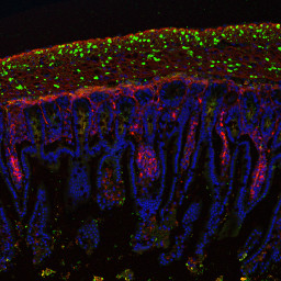
Cross section of intestine of a mouse model of myofibrillar myopathy. In red, alpha smooth muscle actin revealing muscle fibers. In green, mutant desmin causing myopathy, present as aggregates. In blue, the cell nuclei.
Zeiss LSM 700 confocal microscope image of the BFA imaging platform. Dr. Alain Lienbaum and Dr. Eva Cabet.
Oral communication, 10th Euro-IF : European Intermediaire Filaments Meeting , St Malo, France, 14-17 juin 2017
Severe intestinal pseudo-obstruction in a myofibrillar myopathy knock-in mouse model with a R405W desmin mutation.
Cabet E., Delacour D., Hakibilen C., Pichon S., Vicart P., Lilienbaum A

